Diagnosis in digital pathology using whole slide imaging
What is WSI?
Nowadays whole slide imaging(WSI) is changing the world of pathology. WSI is the transition of the traditional microscope. The other name of the WSI is Virtual pathology. In WSI, the tissue slide or glass slide can be scanned and the scanned image is saved into a digital image. Digital image can be share for the discussion and analysis among the doctors and pathology through telepatholoy. Digital images can not spoiled, deteriorate, blur, and break while sharing the images.
What is telepathology?
Telepathology is involved in digital pathology. In which pathologists and doctors communicate through telecommunication technology. Which helps health care providers for collaboration, viewing, analyzing through a mobile and computers. Telepathology technology shrinks the long distance locations for patient care and creates a suitable environment for them in terms of reporting. Diagnosis and treatment.
What is Digital pathology?
Digital pathology provides the digital environment to WSI. Digital pathology is an organization of several pathology, such as Digital pathology association. Which are provide guidelines and tools for digital pathology. In other words, digital pathology focuses on the data management, patient data, image storage that are managed by the digital platform. Digital pathology works on the basis of computer based technology. Digital pathology requires software and hardware. WSI produces a huge data . These types of data is saved on local and cloud-based servers. Digital pathology provides a world-wide network for health care providers. The collection, administration, distribution, and interpretation of pathology data and slides in a digital setting are all included in digital pathology.
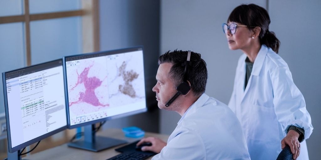
 How does WSI work?
The appropriate scanner is used to capture the images of the biological specimen that are loaded on the glass slide. A thin slice of the tissue is placed on the glass slide along with suitable staining or other protocol for a specific specimen can be scanned and it generates the digitized images. The WSI principle is similar to the traditional microscope, but images by the WSI have more benefits to the pathologist because the pathologist is able to zoom in or zoom out and it has more clarity than images captured by the traditional microscope.
Several types of biological specimens, such as histology samples, cytology slides, micro slides, and hematology smear can be processed using a whole slide image(WSI). WSI has the ability to create high-resolution imaging of tissue specimens and can also produce permanent digital records of slides that enable remote consulting, education, and integrated diagnosis, among other significant advantages.
WSI is also integrated with other digital solutions including Electronic health record(EHR) system or Laboratory information systems(LIS). This connectivity increases or enhances the workflow of the pathologist and enhances patient care. WSI with HER system gives access to the patient . With the help of this, patients can know their previous health history and can share digital slides from the consultant with other doctors. While in the case of LIS, it promotes the laboratory work flow. In which pathologist can view or examine the slide image, related metadata keeps the record in server.

The image of the WSI is displayed on the high resolution monitors. Monitors used in the pathology should have a resolution of 2560*1140 pixels. WSI technology has a Z-stacking facility. Z-stacking is also known as focus stacking. It is a digital imaging method. Which is to gather multiple images at a different focal distance and provide a composite image of the specimen.
AI and ML
Artificial intelligence(AI) and machine learning(ML) technology is effectively used in digital pathology including WSI. This technology identifies the area of interest(ROI) on the pathology slide or classified , computing the cells in the smear. These tools highlight the abnormalities.
Limitations and challenges
- In terms of WSI, there are some limitations and challenges, including infrastructure,organization, integration of other solution software. WSI requires specialized software and equipment .
- organizations who invest in the traditional microscope, it is difficult for them, to invest in the WSI technology on account of maintaining the data or document.
- Image quality is a big challenge for WSI technology. Data management and analysis is also one of the major issues related to the huge volume of data produced by the WSI. And it is also difficult to maintain the security of the patient
Where do we use WSI?
WSI represents the great work flow for research and technology and contributes a novel approach to the pharmaceutical and biotech companies to improve their development and research. WSI is create the digital image library. Because of the high resolution of the image quality, it gives precise information regarding cellular changes of the cells that promote the development.
Objective of WSI in the future
In the world of pathology, the invention of the WSI is the great and novel technology. It creates a digitized image of a glass slide that can be remote to other locations for viewing, analyzing and reporting. In the future, it will make an easy diagnosis of diseases and make the pathology work effortlessly. It keeps the images from being damaged and reduces the financial burden on the patient. It will create the images library for the education, researcher and development . AI and ML are such types of technology integrated with WSI, which contribute to making the pathology world suitable for patients and pathologists.
Reference:
1. https://www.leicabiosystems.com/en-in/life-sciences-and-research-solutions/application/how-whole-slide-imaging-is-changing-pathology/
2. https://developer.nvidia.com/blog/whole-slide-image-analysis-in-real-time-with-monai-and-rapids/
3.https://www.pathologyoutlines.com/topic/informaticstelepathology.html
4.https://scopiolabs.com/ai/introduction-to-ai-in-pathology-main-values-challenges/
5. https://www.pathologynews.com/digital-pathology/whole-slide-imaging-where-does-the-future-lie/


0 comments