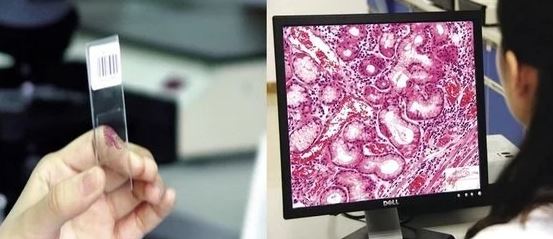
Modernizing Pathology: From Microscope to Digital imaging
The traditional method of pathology reporting involves examining physical glass slides under a light microscope. This approach has been the gold standard for decades, offering high-resolution, direct visualization of tissue samples. It is familiar to most pathologists and does not require sophisticated digital infrastructure. However, it comes with several limitations, including the need for physical storage space, the risk of slide damage or loss, and the difficulty in sharing slides for second opinions or remote consultations, which often requires physically transporting the slides.In the past, examining medical samples (like tissue or blood) depended on highly trained pathologists -doctors who had special education and lots of experience.
However, even though these experts were skilled, manually looking at samples by eye could still be affected by human mistakes likes Cognitive biases (mental shortcuts, expectations, or assumptions that lead to errors). Visual biases (mistakes in how something is seen or interpreted).These biases could cause wrong results or missed problems in the diagnosis.
In contrast, Whole Slide Imaging (WSI) also known as digital pathology or Modernizing pathology which involves scanning entire glass slides into high-resolution digital images that can be viewed on a computer screen. This digital approach significantly enhances efficiency by allowing remote access, easy sharing of slides for consultation, and integration with artificial intelligence tools for image analysis.

Whole Slide Imaging (WSI) offers several important benefits that help pathologists work more efficiently, accurately, and collaboratively. By converting glass slides into high-resolution digital images, WSI enables pathologists to view, analyze, and share image on a computer which makes the modern pathology practice more significant.
Mitigation of Problems occur in pathology after WSI
- WSI greatly improves accessibility. Pathologists can review digital slides anytime and from anywhere, allowing for faster turnaround times and enabling remote work or consultation with colleagues across the globe. This is especially useful in telepathology, where expertise may be needed in locations without a specialist on-site.
- WSI enhances collaboration and second opinions. Since digital slides can be instantly shared, pathologists can easily consult with peers, participate in tumor boards, or contribute to teaching and research with minimal logistical effort.
- WSI allows integration with artificial intelligence and image analysis tools (annotation tool), which can assist in detecting patterns, quantifying features like mitotic figures, and ensuring quality control. This helps reduce human error, improves diagnostic accuracy, and supports more objective reporting.
Additionally, WSI simplifies data management and archiving. Digital slides can be stored, retrieved, and organized much more efficiently than physical slides, reducing the risk of damage or loss and supporting better long-term record keeping.
Overall, WSI empowers pathologists by providing tools for faster diagnosis, easier collaboration, better accuracy, and improved workflow efficiency.Below is flowchart showing the significant changes after the approach of WSI.

Reference:-
1. https://pmc.ncbi.nlm.nih.gov/articles/PMC7522141/
2.https://proscia.com/company/what-is-digital-pathology


0 comments