Histology Department
Histopathology(Histo-tissue / pathos-disease suffering) is one of the branches of the pathology department that deals with the study of types of tissue. The word histopathology is a Greek word. In the histopathology department, the biopsy specimen is taken and examined under a microscope to investigate and study the manifestation of disease or cancer.Histopathology makes diagnosis of disease easy in the medical field and for pathologists for patient care. For the diagnosis of a disease, histopathology refers to microscopy for examination of tissues or surgical specimen.

Why does histopathology perform? Histopatholody is the other branch of diagnosed a disease or cancer. It plays an important role in the medical field. Histopathologiset provides information regarding suspicious diseases or cancer to patients and makes the treatment quickly after they diagnose the condition. Many Histopathologist specialized in many organs, such as the liver, lungs, skin.They take a small tissue from the organ and examine under a microscope. After the specimen is obtained from the patient , the histopathologiest moves toward the further process in the laboratory.
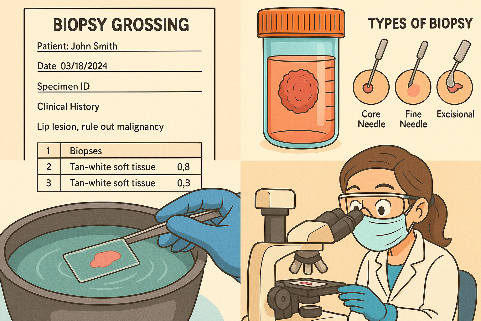
Biopsy: Biopsy is the surgical process of removal of small tissue from the body organs including skin, lungs, liver,other organs or autopsy. To diagnose disease or cancer, biopsy is an accurate procedure along with observing under the microscope. Before performing the staining process, histopathologiest should be fixed, dehydrated, embedded , section, stained, and mounted the specimen. There are several type of biopsy but in the histopathology department, specially focus on the excisional biopsy, incisional biopsy(core biopsy), liquid biopsy and fine needle aspiration biopsy. These biopsies are performed with a microscope.
Excision biopsy: in which the entire lumps or suspicious area is removed.
Incisional biopsy(core biopsy): the specimen is obtained by removing small portion, to avoid removal of whole lumps or tumors.
Liquid biopsy: Liquid biopsy is the laboratory test in which specimens including blood, urine or body fluid are used to detect whether tumour cells are present or not. But liquid biopsy does not give accurate conformation regarding specific cancer ,it only gives small indication regarding cancer. The technological department does not consider that liquid biopsy is a standard method of diagnosing disease or cancer.
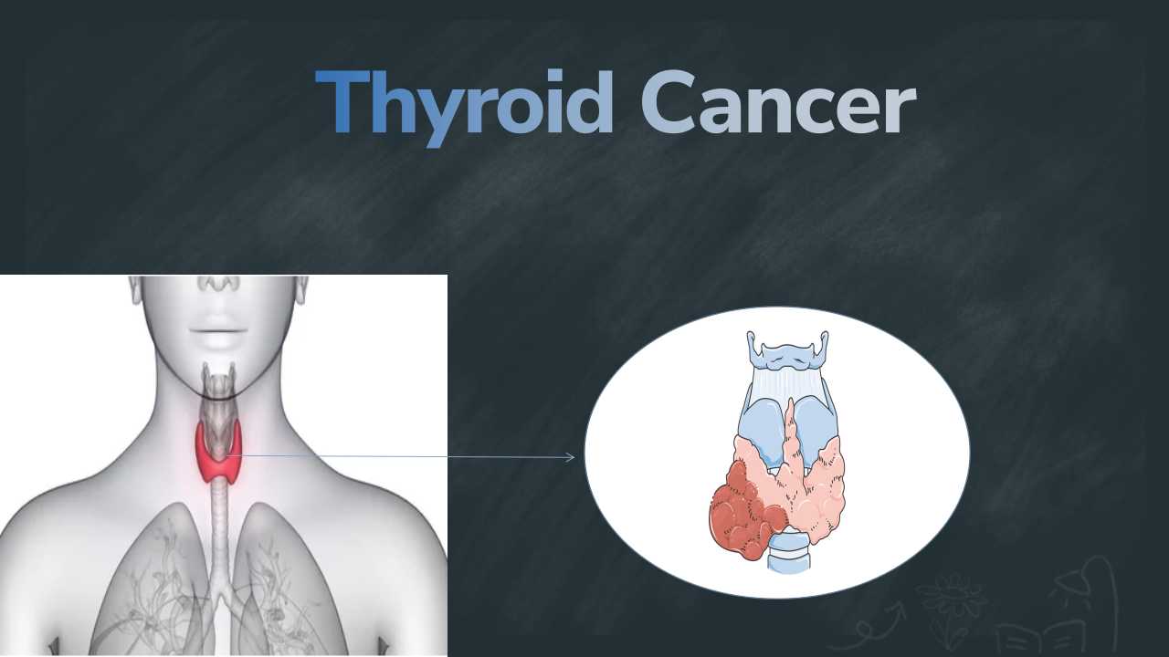
Fine needle aspiration: fine needle aspiration and fine needle biopsy are both the same. In which sharp needle is used to remove the abnormal fluid from the part of the body such as lungs, liver, bone,breast,lymph nodes, thyroid or lesions
The following is the procedure that is performed in the histopathology department after obtaining the specimen from the biopsy of organs and observed under a microscope.

Fixation is an initial step to preparing a specimen to be observed under a microscope. The aim of fixation is to prevent them from decaying.In which the most common fixative is formalin (formaldehyde) , usually in the form of phosphate buffer solution. Formalin stops the enzyme activity, prevents the invading of microorganisms and maintains the molecular structure of tissue to enable or process the staining step.
Grossing is a process in which to keep a record of a patient’s including name, sex, age, medical history. And also keep the records related to specimens, such as type of specimen, number of pieces, and their dimension.
Processing: The specimen are dehyderate (to remove the water) by increase the concentration of alcohol. Xylene is also used instead of alcohol in the late stage of dehydration. Because wax is soluble in xylene and alcohol is not allowed to wax infiltrate. This process is done by an automated instrument is called a “tissue processors”. The wax infiltrated specimen was transferred to the individual specimen embedding container. After this, the molten wax is applied to the specimen in the container and allowed to solidify as embed in the wax block. These steps should be attained carefully and microtome instrument makes thin slice of it for the slide. After cutting the thin slice of wax embedding block, keep it floating on a water-bath surface. To prepare the section for mounted slide.This is an automatic process to preparing a thin slide.
Staining process: The objective of staining is to examine the cellular component of the cells. The cells section stain with different dyes and also use counterstains to provide contrast. The combination of hematoxyline and eosin are mostly used in the histopathology examination to observed under microscope. Hematoxylin is used to stain the nuclei blue, eosin stains the cytoplasm and connective tissue in pink or red color. The other stains also used in histochemistory include seafaring, OilRed O,Congo red, silver stain and artificial dye.
Reference:
1. https://en.wikipedia.org/wiki/Histopathology
2. https://www.prshospital.com/histopathology.php
3. https://www.ncbi.nlm.nih.gov/books/NBK557663/

0 comments