Hypereosinophilic Syndrome
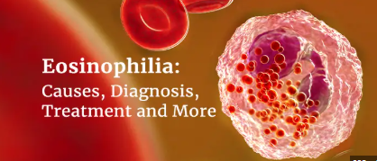
A rare disorder known as hypereosinophilic syndrome (HES) is brought on by an excess of eosinophils, which are white blood cells. Eosinophils perform a number of tasks, such as,protection against infections by parasites as well as against microorganisms inside the cells and also controlling acute hypersensitivity responses. Particularly crucial to the body's defense against parasite infections are eosinophils. T cells secrete hematopoietic growth factors, namely granulocyte-macrophage colony-stimulating factor (GM-CSF), interleukin-3 (IL-3), and interleukin-5 (IL-5), which appear to regulate eosinophil production. IL-5 alone boosts the production of eosinophils, while GM-CSF and IL-3 also stimulate the creation of other myeloid cells.
Typically, white blood cells consist of 5–7% eosinophils, or 100–500 eosinophils per micro-liter of blood. Eosinophils proliferate when people have certain illnesses, like allergies, leading to a condition known as eosinophilia. When eosinophilia quickens, eosinophil synthesis quickens as well, leading to an increase in the quantity of eosinophils, it turns into hypereosinophilic syndrome. This overabundance of eosinophils can harm the heart, lungs, skin, and nervous system, among other organs. Hypereosinophilic syndrome can be fatal if left untreated. Fortunately, over 80% of people with hypereosinophilic syndrome are still living five years following their diagnosis, detection and treatment.
Symptoms
- Skin rashes such as urticaria or angioedema
- Dizziness
- Memory loss or confusion
- Cough
- Shortness of breath
- Fatigue
- Fever
- Mouth sores
Causes/Risk factors
- Genetic factors
- Parasitic infection
- Allergic disorder
- Autoimmune disease
- Hematological disorder
- Lymphoproliferative Disorders
Diagnosis
Hematology department
A peripheral blood smear is prepared on a glass slide by spreading a thin layer of blood. The smear is then fixed with a fixative solution to preserve cell morphology. Giemsa stain is applied to the smear. The staining process with Giemsa stain helps distinguish different cellular components based on their staining properties. Eosinophils, in particular, have distinctive granules that take up the stain, making them easily identifiable under a microscope.After the slide preparation and staining, the slide is ready for the microscopy examination.
Hypereosinophilia under the microscope
Reference:
- https://www.msdmanuals.com/en-in/professional/hematology-and-oncology/eosinophilic-disorders/hypereosinophilic-syndrome#v9877132
- https://my.clevelandclinic.org/health/diseases/22541-hypereosinophilic-syndrome


0 comments