Leishmaniasis
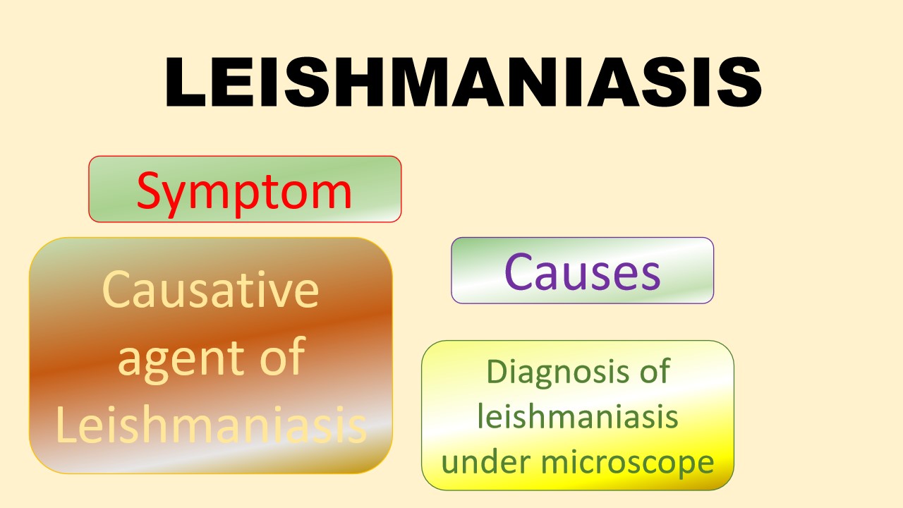
The Leishmania parasite causes the parasitic disease called Leishmaniasis. Usually, this parasite inhabits infected sand flies. If an infected sand fly bites you, you could get leishmaniasis.Through the bite of the phlebotomine sand fly, leishmaniasis is transmitted. After biting an animal or person who is infected, the sand fly injects the parasite into another. These types of sand flies are found in the tropical and subtropical environment. Once upon a time, this disease was fetal epidemic in the area of Asia, East Africa, and South America because of shortage or limited source of treating disease.
Cutaneous, visceral, and mucocutaneous, these are the types of leishmaniasis. Each form of leishmaniasis is associated with specific species of leishmania parasite. Cutaneous leishmaniasis, this is most common type of leishmaniasis. It affects the skin and causes ulcers. Mucocutaneous leishmaniasis, it is dangerous types of leishmaniasis. In which the parasite spreads to the nose, throat, and mouth. In this area, the parasite destroys the mucous membranes. Mucocutaneous leishmaniasis is the result of the cutaneous leishmaniasis. Visceralleishmaniasis, it is the serious form disease. Visceral leishmaniasis is also called Kala-azar. In which leishmania affect the internal organ such as spleen and liver and damage it.
Symptoms
- painless skin ulcers
- runny or stuffy nose
- nosebleeds
- difficulty breathing
- weakness
- fever that lasts for weeks or months
- enlarged spleen
- enlarged liver
- decreased production of blood cells
- swollen lymph nodes
Causes/Risk factors
- Transmission through Sandfly Bites
- Reservoir Hosts (rodents, dogs, or other mammals)
- Vector-Borne Transmission (sand-flies of the genus Phlebotomus)
- Geographic Distribution
Diagnosis
Histology examination:
The bone marrow specimen is checked under the microscope for diagnosis of leismaniasis.
Bone marrow specimen
Microbiology examination:
The culture method is performed then colonies spread on the slide , stain it and checked under the microscope.
Microbiology examination
Cytology Department:
the skin lesion can be performed for histopathological examination to diagnose the leishmaniasis. To prepare the microscopy slide with the specimen.This includes examining tissue samples under a microscope to see whether Leishmania parasites are present or not and to evaluate any tissue damage. The slide was stained with the Giemsa stain and examined under the microscope.
Leishmania tropica under the microscope
References:
1.https://my.clevelandclinic.org/health/diseases/24539-leishmaniasis
2.https://www.who.int/news-room/fact-sheets/detail/leishmaniasis


0 comments