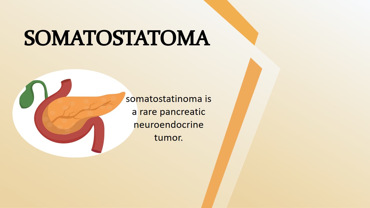
Somatostatinoma
Somatostatinoma is one of the types of neuroendocrine tumor of pancreas. Pancreas is vital part of the digestive system of the body. Based on the function of body, pancreas divided into two part exocrine pancreas and endocrine pancreas. Exocrine pancreas which produce digestive enzyme and Endocrine pancreas which are responsible for producing insulin, glucagon,somatostatino hormones. there are the five types of the pancreatic neuroendocrine tumor. Which are called insulinoma, gastrinoma, somatostatinomas, glucagonoma and VIpoma. Neuroendocrine tumor is a cancer, that are begin in the neuroendocrine cells or organs. What is neuroendocrine cells? Neuroendocrine cells make hormones and release them into the blood stream. Neuroendocrine cells are present in most of organ in the body include esophagous, stomach, lungs, small and large bowel, pancreas liver.
Somatostatinoma is a rare cancer of neuroendocrine tumor of pancreas , develops in the head of the pancreas and duodenum. In the Somatostatinomas disease, hormone like somatostatin are excessively produce. The excessive production of somatostatino inhibits the release of other hormones. Which are important for other functions of the body. The most common clinical manifestation of somatostatinoma is stomach pain and gradually weight loss.
Symptms
- loss of appetite
- Nausea and vomiting
- abdominal pain
- frequent diarrhea
- unintentional weight loss
- Gallstones
- Fatty poo (stools)
- Jaundice
- Blockage in the bowel
Causes/Risk factor
- Smoking
- Drinking alcohol
- Family history of cancer
- Pancreatitis
- Hereditary or with other rare genetic syndromes(neurofibromatosis endocrine neoplasia type 1, and Von Hippel-Lindau syndrome)
- overweight and obesity
- chronic pancreatitis
- stomach infection
Diagnosis
Cytology Department: The Fine needle aspiration(FNA) cytology method is used for somatostatinomas diagnosis under a microscope.The specimen is collected from the liver(Ascites) and sent to the laboratory. The pathologists make smear and stain the hematoxylin and eosin. Under the microscope to observe the smear have more cells arranged in loosely cohesive groups.Hematoxylin, which stains the cells' nuclei blue, and Eosin, which stains the extracellular matrix and cytoplasm in pink. This depends on which method they use.

FNS cytology smear stain with hematoxylin and eosin observe under the microscope show the large number of cells
Histology Department:
The biopsy of the pancreatic tissue observed under the microscope. The tissue stained with hematoxylin and eosin stain.  A malignant metastasizing PNT located in the tail of the pancreas
Reference:
1. https://www.medicalnewstoday.com/articles/somatostatinomas#seeing-a-doctor
2. https://www.dovemed.com/diseases-conditions/somatostatinoma


0 comments